Radiology Department

Offering a comprehensive range of sophisticated imaging, The CDUH Department of Radiology is equipped with up-to-date digital technology while maintaining a welcoming environment that focuses on quality and patient comfort. Several recent enhancements are in place to ensure efficient and accurate tests for easy diagnosis.

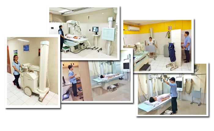
FACILITIES AND SERVICES

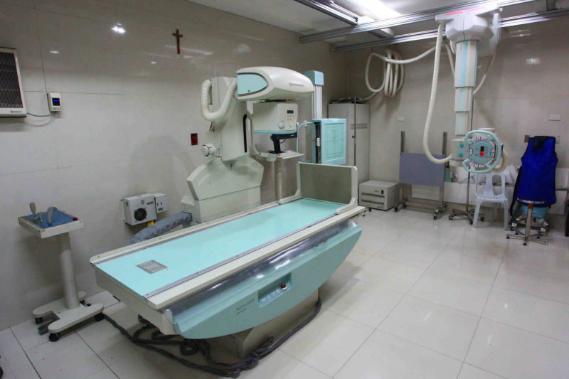
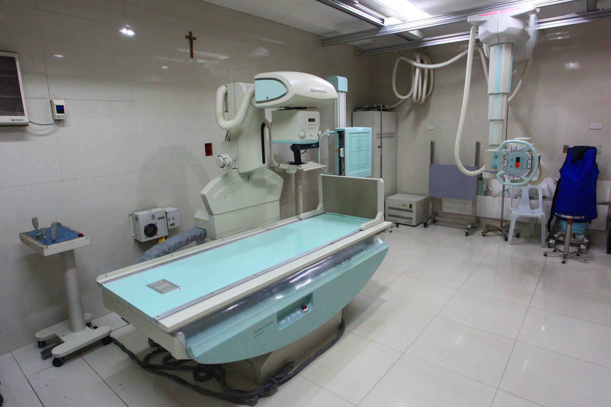
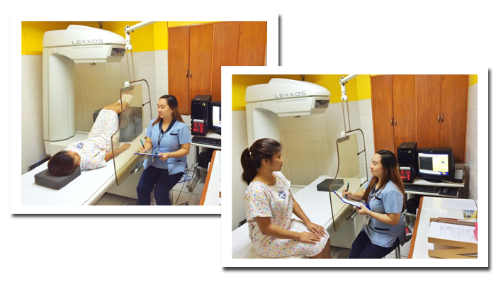
A bone density test is a measurement of the density of your bone mineral, and can be taken from various places throughout the body. A bone density test involves the use of x-ray, in low dosage, and takes only few minutes with virtually no discomfort to the patient. The bone density test is the signle most useful procedure in the diagnosis of osteoporosis and the evaluation of fracture risk. The lower your bone density, the higher your risk of fracture.

BMD

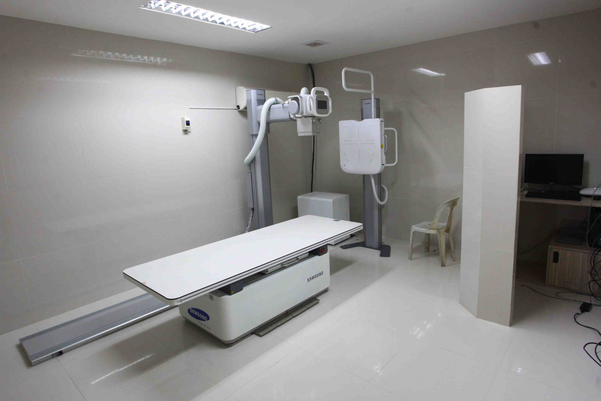
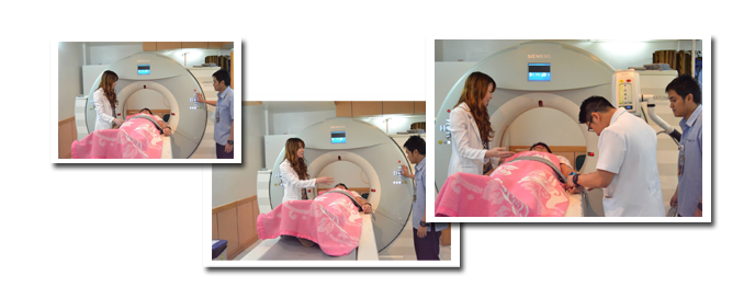
It uses X-rays to create numerous cross-sectional images of the body. CT is used to diagnose disease and injury in many parts of the body such as trauma, oncology, gastrointestinal care, postoperative evaluation, chest disorders, orthopedics, and angiographies. CDUH's advance CT imaging systems (Siemens Definition AS+ 128-Slice),with precise and rapid imaging of the body, provide highly detailed three-dimensional images which can be used to diagnose common and hard-to-detect conditions in areas such as the head (stroke assessment), neck, chest, heart and blood vessels, abdomen, lungs, colon, and legs.

CT

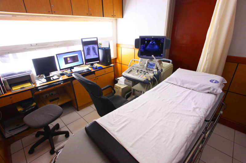
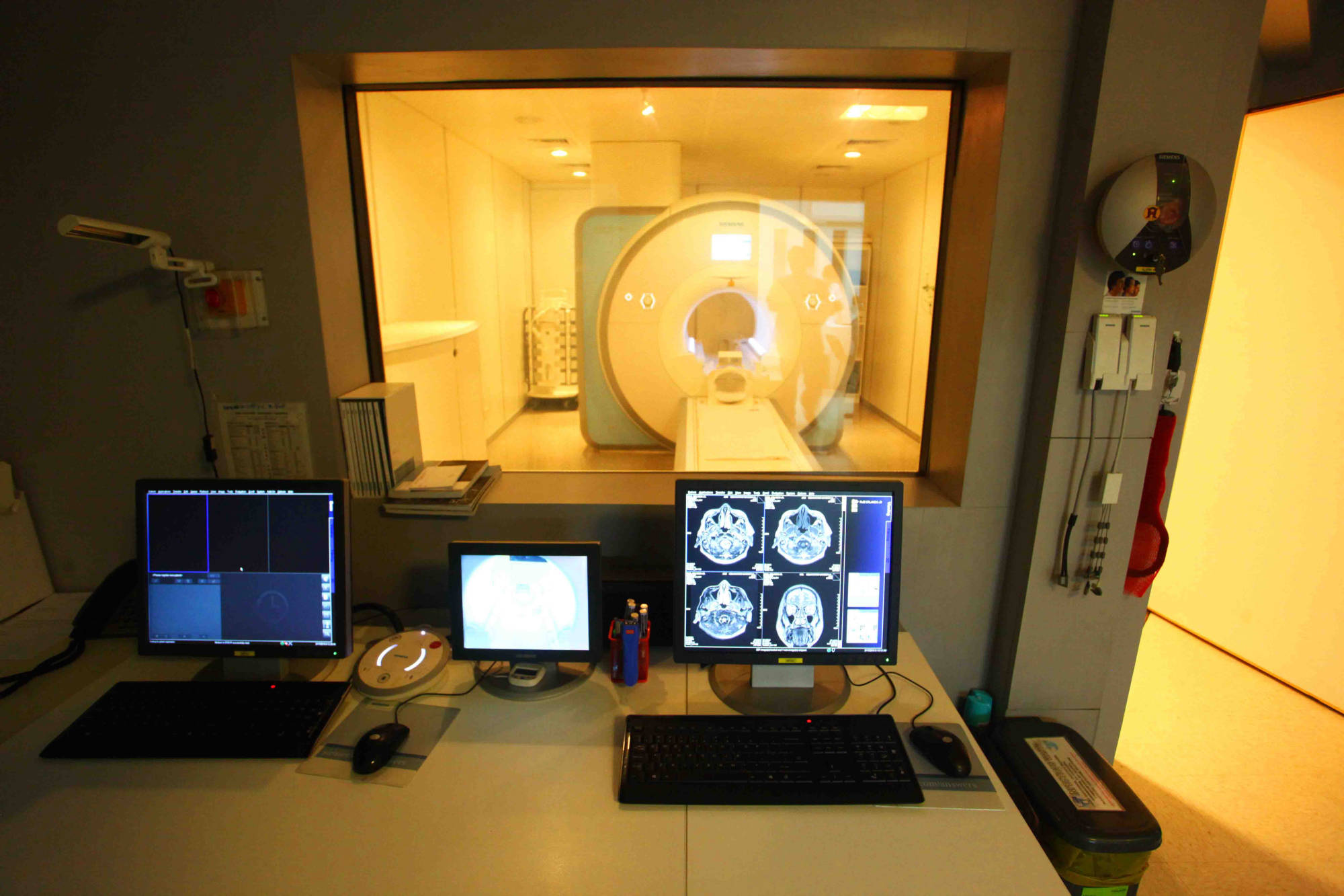
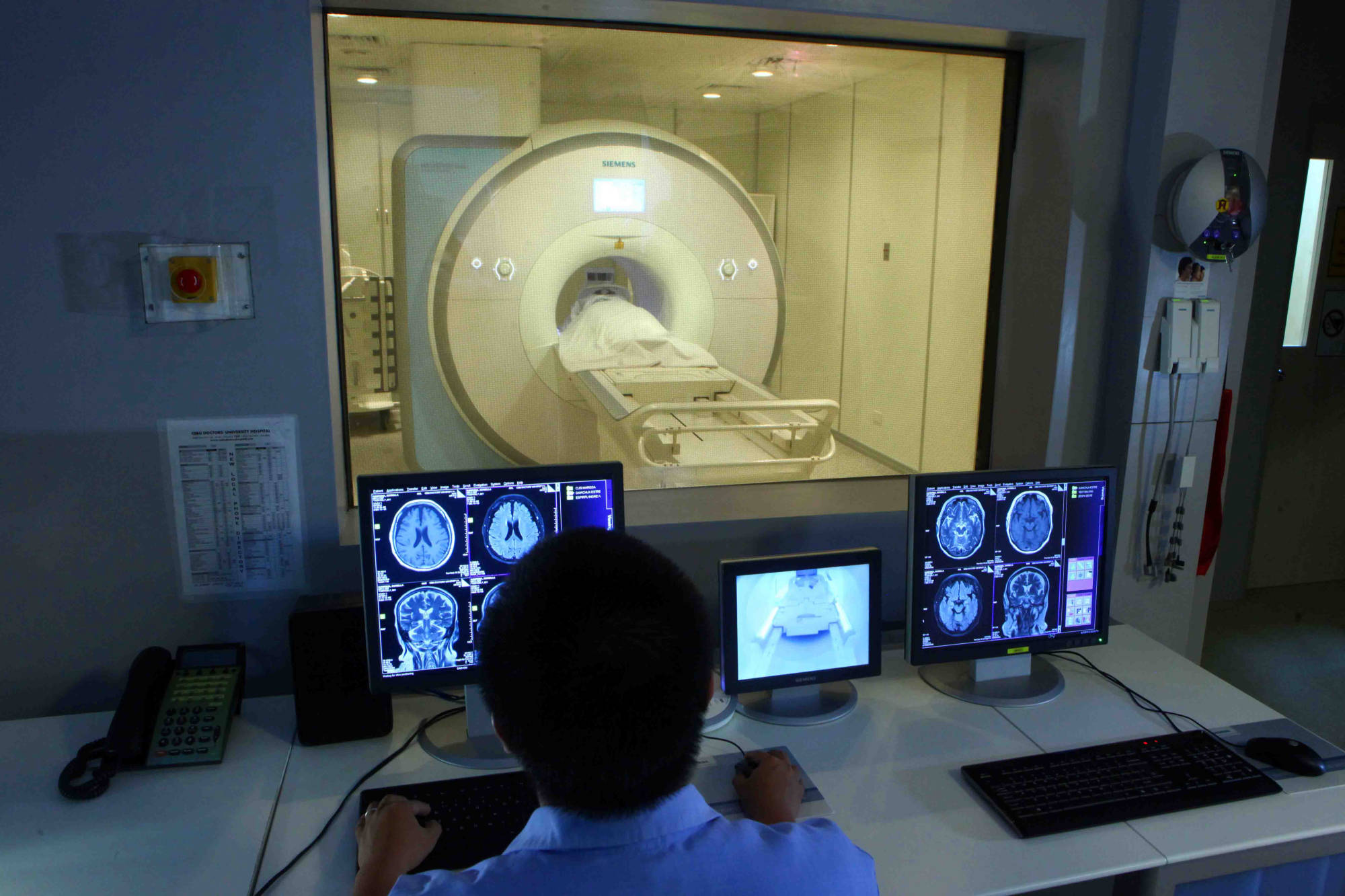
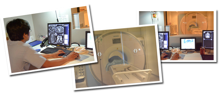
An MRI (magnetic resonance imaging) scan is a radiology technique that uses magnetism, radio waves, and a computer to produce images of body structures. The MRI scanner is a tube surrounded by a giant circular magnet. The patient is placed on a moveable bed that is inserted into the magnet. The magnet creates a strong magnetic field that aligns the protons of hydrogen atoms, which are then exposed to a beam of radio waves. This spins the various protons of the body, and they produce a faint signal that is detected by the receiver portion of the MRI scanner. The receiver information is processed by a computer, and an image is produced.

MRI SCAN FACTS:

  • MRI scanning uses magnetism, radio waves, and a computer to produce images of body structures.
  • MRI scanning is painless and does not involve x-ray radiation
  • Patients with heart pacemakers, metal implants, or metal chips or clips in or around the eyes cannot be scanned with MRI because of the effect of the magnet.
  • Claustrophobic sensation can occur with MRI scanning.
MRI

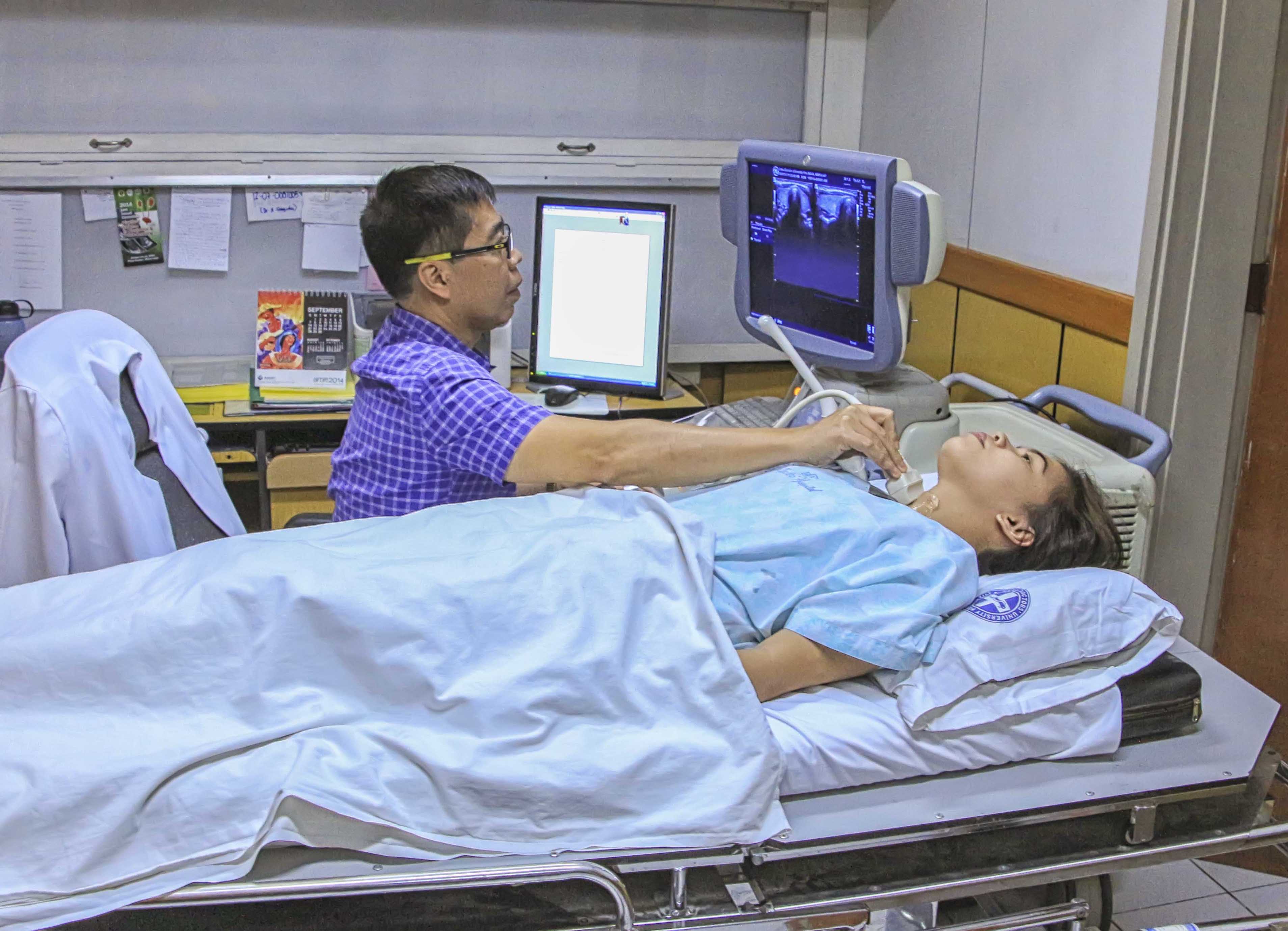
It is safe and painless, and produces pictures of the inside of the body using sound waves. Ultrasound imaging, also called ultrasound scanning or sonography, involves the use of a small transducer (probe) and ultrasound gel placed directly on the skin. High-frequency sound waves are transmitted from the probe through the gel into the body. The transducer collects the sounds that bounce back and a computer then uses those sound waves to create an image. Ultrasound examinations do not use ionizing radiation (as used in x-rays), thus there is no radiation exposure to the patient. Because ultrasound images are captured in real-time, they can show the structure and movement of the body's internal organs, as well as blood flowing through blood vessels.

ULTRASOUND

OTHER SERVICES OFFERED

  • ULTRASOUND IMAGING
  • COMPUTED TOMOGRAPHY SCAN ( CT-SCAN)
  • DIGITAL X-RAY

The MRI Section is equipped with the most advanced Siemens Magnetum Aera, designed to make exams easier for the radiographer and more comfortable for the patients.

The Ultrasound Imaging or Sonography has high-definition and enhanced diagnostic capabilities with their GE Logiq S6.

The digital X-ray section with four rooms for both in and outpatients is using the latest Samsung GF50 which guarantees the best quality of images and accurate diagnosis.

The Bone Mass Densitometry section assesses the strength of bones and probability of fractures for those with the risk of osteoporosis.

The CT-SCAN diagnosis the diseases and injuries in various parts of the body. The state-of-the-art GE Light Speed VCT 64-Slice and Siemens Definition As + 128-Slice creates precise and rapid images of the body for the best possible scans.

FAQ BMD (BONE MINERAL DENSITOMETRY)

Is a condition characterized by low bone density, which simply means there is less bone compared to when you were young and your bones were at their peak, healthy stage. Because there is less bone density, the bones become brittle, weak and more likely to fracture.

Certain factors are linked to the development of osteoporosis or contribute to the likelihood of developing the condition. These are called risk factors. Your physician may recommend a bone density test if:

  1. You have gone through menopause, or
  2. You have major risk factor for osteoporosis such as menopause before the age of 45, or
  3. You have family history of osteoporosis.

Because bone density test is safe, simple and quick, there are no special preparations necessary for density tst.

If you are pregnant, you should not undergo any x-ray procedure unless advised otherwise by your physician.

FAQ MRI (MAGNETIC RESONANCE IMAGING)

MRI is done for many reasons. It is used to find problems such as tumors, bleeding, injury, blood vessel diseases, or infection. MRI also may be done to provide more information about a problem seen on an X-Ray, Ultrasound scan, or CT Scan. Contrast material may be used during MRI to show abnormal tissue more clearly. An MRI scan can be done for the:

  • Head. MRI can look at the brain for tumors, an aneursym, bleeding in the brain, nerve injury, and other problems, such as damage caused by a stroke. MRI can also find problems of the eyes and optic nerves and the ears and auditory nerves.
  • Chest. MRI of the chest can look at the heart, the valves, and coronary blood vessels. It can show if the heart or lungs are damaged. MRI of the chest may also be used to look for breast or lung cancer.
  • Blood Vessels. Using MRI to look at blood vessels and the flow of blood through them is called Magnetic Resonance Angiography (MRA). It can find problems of the arteries and veins, such as an aneurysm, a blocked blood vessel, or the torn lining of a blood vessel (dissection). Sometimes contrast material is used to see the blood vessels more clearly.
  • Abdomen and Pelvis. MRI can find problems in the organs and structures in the belly, such as the liver, gallbladder, pancreas, kidneys, and bladder. It is used to find tumors, bleeding, infection, and blockage. In women, it can look at the uterus and ovaries. In men, it looks at the prostate.
  • Bones and joints. MRI can check for problems of the bones and joints, such as arthritis, problems with the temporomandibular joint, bone marrow problems, bone tumors, cartilage problems, torn ligaments or tendons, or infection.
  • Spine. MRI can check the discs and nerves of the spine for conditions such as spinal stenosis, disc bulges, and spinal tumors

Patients who have any metallic materials within the body must notify their physician prior to the examination or inform the MRI staff. Metallic chips, materials, surgical clips, or foreign material (artificial joints, metallic bone plates, or prosthetic devices, etc.) can significantly distort the images obtained by the MRI scanner. Patients who have heart pacemakers, metal implants, or metal chips or clips in or around the eyeballs cannot be scanned with an MRI because of the risk that the magnet may move the metal in these areas. Similarly, patients with artificial heart valves, metallic ear implants, bullet fragments, and chemotherapy or insulin pumps should not have MRI scanning.

All metallic objects on the body are removed prior to obtaining an MRI scan. MRI scanning requires that the patient lie still for best accuracy. Patients lie within a closed environment inside the magnetic machine. Relaxation is important during the procedure and patients are asked to breathe normally. Interactin with the MRI technologist is maintained throughout the test. There are loud, repetitive clicking noises which occur during the test as the scanning proceeds. Occasionally, patients require injections of liquid intravenously to enhance the images which are obtained. The MRI scanning time depends on the exact area of the body studied, but ranges from half an hour to an hour and a half.

FAQ ULTRASOUND

Ultrasound is a useful way of examining many of the body's internal organs, including but not limited to the:

  • heart and blood vessels, including the abdominal aorta and its major branches
  • liver
  • gallbladder
  • spleen
  • pancreas
  • kidneys
  • bladder
  • uterus, ovaries
  • eyes
  • thyroid and parathyroid glands
  • scrotum (testicles)
  • brain in infants
  • hips in infants

You should wear comfortable, loose-fitting clothing for your ultrasound exam. You may need to remove all clothing and jewelry in the area to be examined. You may be asked to wear a gown during the procedure. Preparation for the procedure will depend on the type of examination you will have. For some scans your doctor may instruct you not to eat or drink for as many as 12 hours before appointment. For others you may be asked to drink up to six glasses of water two hours prior to your exam and avoid urinating so that your bladder is full when the scan begins.

BENEFITS

  • Most ultrasound scanning is noninvasive (no needles or injections).
  • Occasionally, an ultrasound exam may be temporarily uncomfortable, but it is almost never painful.
  • Ultrasound is widely available, easy-to-use and less expensive than other imaging methods.
  • Ultrasound imaging is extremely safe and does not use any ionizing radiation.
  • Ultrasound scanning gives a clear picture of soft tissues that do not show up well on x-ray images.
  • Ultrasound is the preffered imaging modality for the diagnosis and monitoring of pregnant women and their unborn babies.
  • Ultrasound provides real-time imaging, making it a good tool for guiding minimally invasive procedures such as needle biopsies and fluid aspiration.

RISKS

  • For standard diagnostic ultrasound, there are no known harmful effects on humans.

RADIOLOGY DEPARTMENT
G/F Cebu Doctors' University Hospital
Tel. #: (+63) 32 255 - 5555

CT-Scan - Local 110
MRI - Local 127
Ultrasound - Local 126
X-Ray - Local 125


cebudoc
Osmeña Blvd., Cebu City
+63 032 255-5555

mactandoc
Basak, Lapu-lapu City
+63 032 2397002 to 7016

northgen
Talamban, Cebu City
+63 032 343-7777

sancarlosdoc
San Carlos City, Negros Occidental
+63 034 312-5136

southgen
City of Naga, Cebu
+63 032 272-2223

ormocdoc
Ormoc City, Leyte
+63 053 255-7522

Contacts

Email: info@cebudocgroup.com.ph
Phone: 032 255-5555

Feedback

Please send us your ideas, bug reports, suggestions! Any feedback would be appreciated.